Taking molecular pathology to the next level: Whole slide multicolor confocal imaging with the Pannoramic Confocal digital pathology scanner
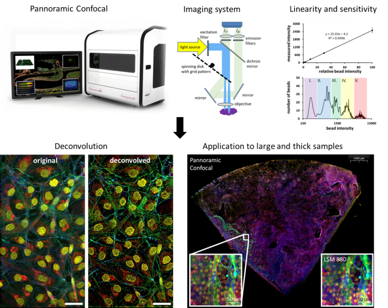
The Pannoramic Confocal (created by 3DHistech, Ltd. Hungary) is a first-of-the-kind digital pathology scanner that affords not only multiplexed fluorescent detection on top of conventional transmission imaging, but also confocality. We have benchmarked and optimized, together with 3DHistech Ltd, this scanner in terms of stability, precision, light efficiency, linearity, and sensitivity within the project GINOP-2.2.1-15-2017-00072. X-Y stability and relocalisation precision were well below resolution limit (≤50 nm). Light throughput in confocal mode was 4-5 times higher than that of a point scanning confocal microscope, yielding similar calculated confocal intensities but with the potential for improving signal to noise ratio or scan speed. Response was linear with R² ≥0.9996. Calibrated measurements showed that using indirect labeling ≥ 2000 molecules per cell could be well detected and imaged on the cell surface. Both standard-based and statistical post-acquisition flatfield corrections were implemented. The dimensions of the PSF were somewhat larger and less symmetric than of the theoretical PSF of a conventional CLSM, however, the spatial homogeneity of these parameters allowed for obtaining a specific system PSF for each optical path and using it for optional on-the-fly deconvolution. In conclusion, the Pannoramic Confocal provides sensitive, quantitative widefield and confocal detection of multiplexed fluorescence signals, with optical sectioning and 3D reconstruction, in addition to brightfield transmission imaging. High speed scanning of large samples, analysis of tissue heterogeneity, and detection of rare events open new ways for quantitatively analyzing tissue sections, organoid cultures, or large numbers of adherent cells.
Rebenku István, Bartha A. Ferenc, Katona Tamás, Zsebik Barbara, Antalffy Géza, Takács Lili, Molnár Béla, Vereb György
https://doi.org/10.1002/cyto.a.24675