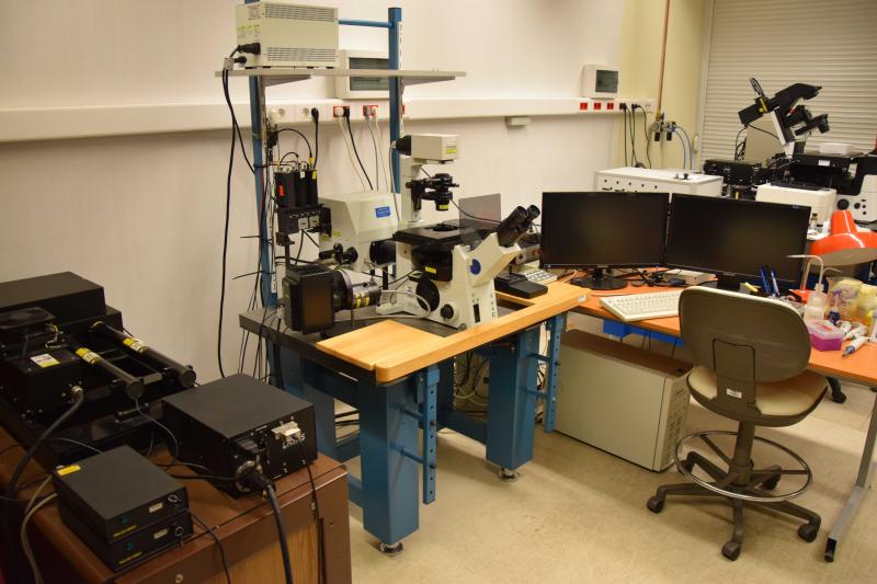
Name of the system: OLYMPUS FluoView FV1000 Confocal Microscope
Location of the instrument: Life Science Building 2nd floor; 2.045 Room
Organisation: Sándor Damjanovich Cell Analysis Core Facility, Department of Biophysics and Cell Biology, Faculty of Medicine, University of Debrecen
Core Facility Homepage: biophys.med.unideb.hu/en/cellularimaging E-mail: dszl@med.unideb.hu
Registration:
- Only authorized and trained Users are allowed to use the Equipment in the Core Facility!
- Users need to attend an instrument specific training program before being allowed to sign up to use the equipment independently.
Online instrument booking: link
Specifications:
Olympus FluoView 1000 confocal laser scanning microscope and fluorescence correlation spectroscope. The 3 lasers of the microscope provide excitation wavelengths of 456, 488, 514, 543 and 633 nm. 3 fluorescence signals - of which 2 with spectral resolution - and one transmitted light signal (with DIC contrast) can be detected from the sample at the same time. Equipment suitable for microscopic tomography and detection of molecular mobility and co-movement. By scanning, images with an optical slice thickness of 0.5 μm can be achieved, from which the 3D image of the sample, the spatial distribution and colocalization of the biomolecules can be reconstructed. A uniquely designed, three-channel fluorescence correlation and cross-correlation spectroscope (FCS, FCCS) is connected to the microscope.
Potential use:
- co-localization analyses with 200 nm resolution
- multichannel confocal imaging
- multichannel fluorescent correlation and cross-correlation spectroscopy
- optical sectioning, 3D reconstruction
- FRET, FRAP measurements